Attic Retraction Pocket Cases
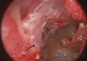
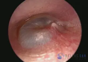
Attic Retraction Pocket Cholesteatoma Case 1
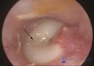
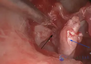
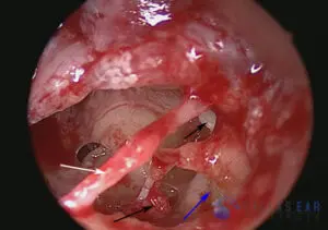
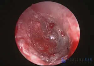
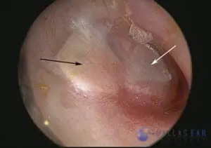
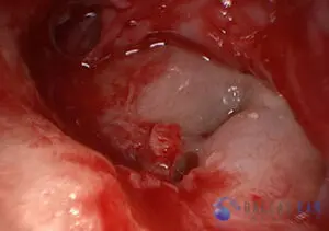
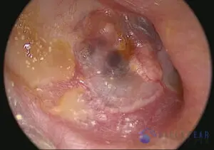
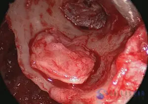
Attic retraction pocket cholesteatoma is clearly visualized (white arrow). There has been significant bone erosion of the ear canal wall above the eardrum. Skin material often accumulates in this pocket and becomes infected, causing drainage and potential severe complications. The remainder of the eardrum shows some myringosclerosis (blue arrow), or scarring of the earfdrum, from a history of chronic infections.
Attic Cholesteatoma Case 1
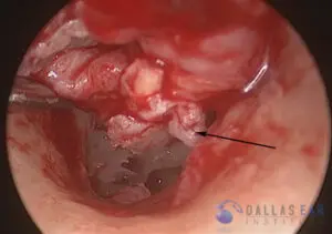
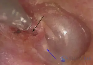
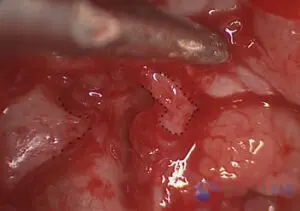
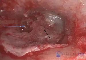
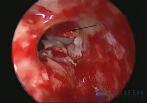
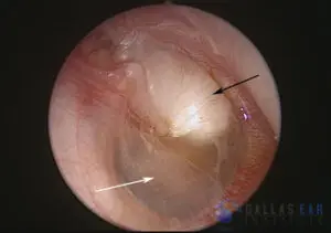
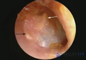
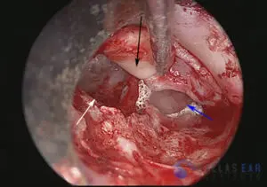
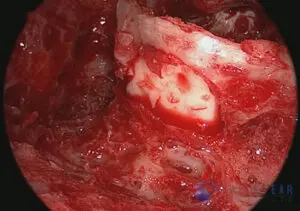
- The tympanic membrane (eardrum) is visualized through the ear canal. The defect in the ear drum is seen and indicated with the black arrow. This is a cholesteatoma that has formed. The area of the superior portion of the eardrum is retracted, or sucked in, trapping skin cells and debris and eating away at the hearing bones and ear canal bone. The blue arrow shows the cholesteatoma pocket within the middle ear.

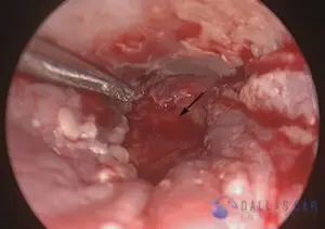
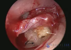
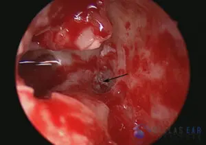
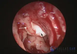
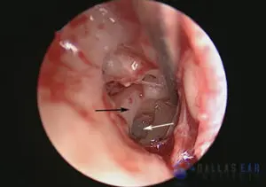
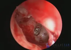
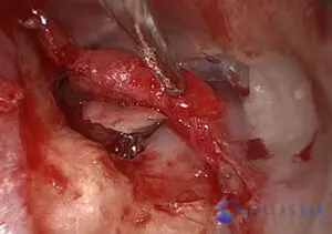
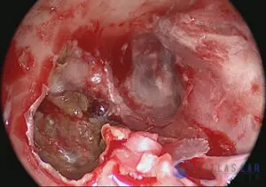
- The eardrum has been lifted by making an incision in the ear canal. There is significant inflammation in the middle ear as well as cholesteatoma sitting on top of, and eroding into, the hearing bones (black arrow).
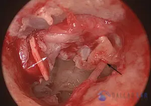
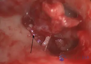
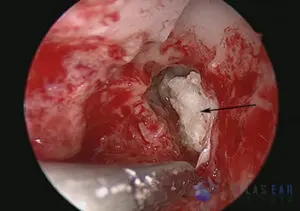
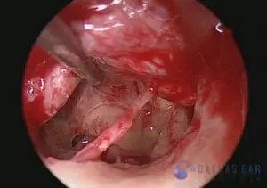
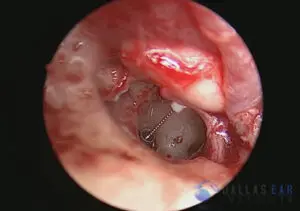
- The blue arrow is showing the cholesteatoma within the middle ear cavity. This skin accumulates and damages the structures of the middle ear and inner ear. The black arrow is pointing to an eroded section of the incus, the 2nd hearing bone, that has been eaten away by the cholesteatoma.
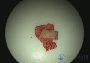
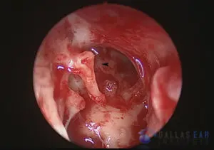
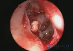
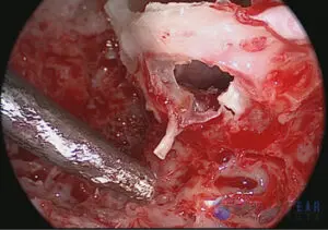
- This image shows the remnant of the 2nd hearing bone, the incus, commonly known as the anvil, that has been eaten away. This causes a significant hearing loss.
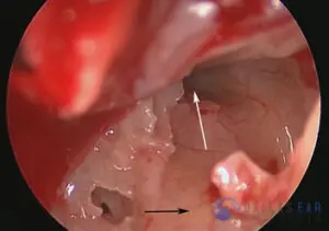
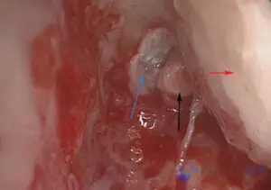
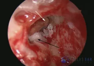
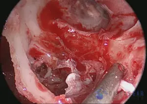
- A view through the mastoid cavity, which is behind and above the ear canal, is seen. The red arrow signifies the ear canal bone, which is intact. The black arrow represents the head of the incus, the 2nd hearing bone. The blue arrow is pointing towards significant skin elements within the upper part of the middle ear and opening into the mastoid. This trapped skin signifies the cholesteatoma.
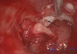
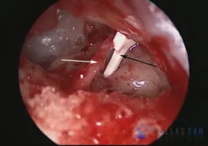
- The malleus, or 1st hearing bone is clearly seen now that the eardrum has been lifted off of it. The blue arrow is showing the stapes bone, the 3rd hearing bone, that conducts sound into the inner ear. The black arrow is showing the undersurface of the remnant eardrum.
- The reconstruction of the defect that the cholesteatoma caused is prepared. A cartilage and perichondrail graft is utilized to rebuild the ear canal bone that has been eaten away by the cholesteatoma. This will repair this defect and prevent future retraction.
- The remnant eardrum is indicated by the black arrow. The defect in the eardrum and ear canal wall is shown with the blue arrow. Reconstruction of this defect is required.
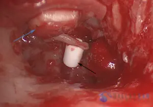
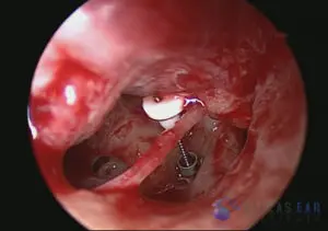
- Because the 2nd hearing bone was eroded by the cholesteatoma, the hearing bones must be reconstructed to rebuild the hearing mechanism. Thus, in this case, a hydroxyapatite prosthesis is used to bridge the gap between the 1st hearing bone and the 3rd hearing bone. This tubular structure sits atop the stapes, the 3rd hearing bone, and connects this bone to the malleus, the 1st hearing bone. Thus, when sound hits the eardrum, it will vibrate the fluid of the inner ear through this mechanism. In addition, the cartilage graft is seen (blue arrow) from the undersurface and will repair the ear canal defect.
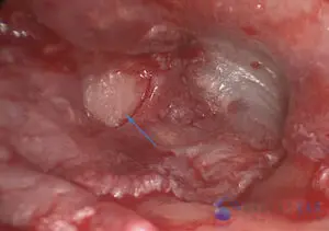
- The final repair from the ear canal is now visualized. The cartilage repair is seen and is in excellent position for expected healing.
Congenital Cases
Cholesteatoma - Congenital Case 1
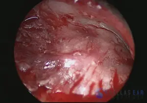
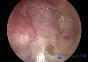
- A photograph of the eardrum. This eardrum is normal appearing. There is no evidence of previous perforation and there are no retraction pockets.
- After lifting up the eardrum, frank cholesteatoma is noted within the middle ear cavity (white arrow). The black arrow is pointing towards the round window niche of the inner ear. As the cholesteatoma does not originate from the eardrum skin, it is termed a congenital cholesteatoma. This means that it was present at birth and is not a result of chronic ear problems.
- Mastoidectomy has been performed. When approaching the interface between the mastoid cavity and the middle ear cavity, cholesteatoma is noted to be filling this area (black arrow). This cholesteatoma is lining the bone that separates the ear and mastoid from the brain, but has not eroded through this bone.
- Another view from the mastoid cavity into the uppermost region of the middle ear cavity. The white arrow points toward the head of the incus, the second hearing bone. Cholesteatoma is clearly seen (black arrow), and is directly on top of the bony canal that holds the facial nerve. This bone overlying the facial nerve may be eroded by cholesteatoma when left untreated for a long period.
- A view of the middle ear cavity after much of the cholesteatoma has been removed. Note cholesteatoma (black arrows) that is surrounding the stapes bone and also is present in the anterior middle ear space. In addition, the blue arrow points toward the cholesteatoma that is present on top of the facial nerve. The white arrow signifies the chorda tympani taste nerve.
- The incus has been removed, and here we see the head of the malleus that lies just in front of the incus (black arrow). The cholesteatoma continues to be removed.
- Cholesteatoma matrix has grown into a petrous apex air cell toward the midline of the skull (black arrow).
- A view of the middle ear cavity after all cholesteatoma has been removed.
- After confirmation of complete cholesteatoma removal, the hearing is reconstructed. In this patient, a titanium shoe is placed on the footplate to the inner ear and a prosthesis is used to bridge the gap between the malleus (first hearing bone) and the shoe on the footplate of the inner ear. A small cartilage graft is placed between the eardrum and the head of the prosthesis. The hearing mechanism is thus re-established.
Cholesteatoma - Congenital Case 2
- A child with a whitish mass seen in the middle ear, behind the eardrum. The eardrum is intact with no visible retraction pockets and no previous history of perforations (potential causes of cholesteatoma). The congenital cholesteatoma (black arrow) is in front of the malleus, the first hearing bone, and is pressing up on the eardrum itself. This disease is not caused by chronic ear infections, but occurs in individuals that are born with skin cells trapped behind an intact eardrum. The posterior, or back part, of the eardrum and middle ear appears to be free of cholesteatoma (white arrow).
- After lifting up the eardrum, the cholesteatoma is clearly visualized. The eardrum has been carefully lifted off of the malleus (white arrow). The cholesteatoma (black arrow) has no attachment to the undersurface of the eardrum. This congenital cholesteatoma was easily removed.
- The area where the cholesteatoma sat is now free of disease (black arrowhead). All skin elements have been removed from the middle ear cavity. The hearing bones, or ossicles, have not been damaged by the cholesteatoma.
- The eardrum, or tympanic membrane, is set back into place. The eardrum was completely intact with no involvement by the cholesteatoma and no damage during the surgery.
2nd Look at Middle Ear Cases
2nd Look Middle Ear Cholesteatoma Case 1
- A view of the reconstructed eardrum. This patient had an extensive cholesteatoma with significant erosion of the ear canal bone and ossicles that was surgically resected 6 months previously. The patient was taken for middle ear exploration to ensure no residual disease and to reconstruct the eardrum.
The black arrow indicates the previously placed cartilage graft, which is directly underneath the eardrum and recreates the ear canal wall that was eroded by cholesteatoma and drilled for adequate visualization of the disease process during the original ear surgery. The white arrow indicates the first hearing bone which is immediately underneath the eardrum—the malleus. This bone is nearly always maintained in the middle ear during surgery, unless involved with cholesteatoma. - View of the middle ear cavity after eardrum has been lifted. The stapes footplate is indicated by the white arrow. This is the thin shell of bone that separates the inner ear cavity from the middle ear cavity. In a normal working ear, there are three bones that reside in the middle ear cavity, and the stapes bone, or stirrup bone, sits on this footplate and vibrates the fluid within the inner ear to allow us to hear. Because this patient had extensive erosion of this bone, along with the 2nd hearing bone, the incus, these are no longer present. The black arrow points to the horizontal portion of the facial nerve, the nerve that allows all facial movement. This nerve lies in a bony tunnel just above the stapes footplate.
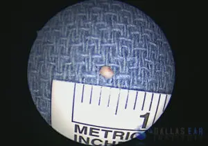
- Small cholesteatoma pearl in middle ear cavity. This tiny pearl, or circumscribed skin ball, was found during the 2nd look procedure. In this photo, the tiny pearl was seen under the ear canal bone and was dissected carefully and removed in whole. Often, if some skin cells are hidden from view and unable to be removed during the primary surgery, they form into a pearl such as this which is easily and completely removed during the 2nd look procedure, with little to no chance of residual disease.
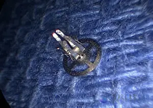
- A view of the tiny cholesteatoma pearl that was removed from the middle ear cavity. Note the size of the skin pearl. The ruler adjacent to the skin pearl indicates that it’s size is less than 2 mm.
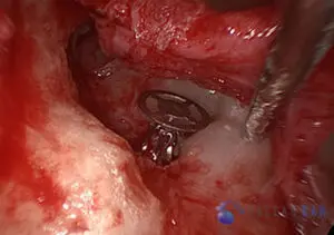
- Hearing reconstruction. Once any disease has been removed from the middle ear cavity, the hearing mechanism is then rebuilt, first by placing a shoe over the footplate. This titanium shoe accepts the ossicular prostheses and allows re-establishment of the hearing mechanism.
- Hearing reconstruction. The titanium prosthesis with a hydroxyapatite head is then placed from the footplate shoe to the malleus on the undersurface of the eardrum. The connection between the eardrum and the inner ear is re-established.
2nd Look Middle Ear Cholesteatoma Case Number 2
- View of eardrum and cartilage graft. Patient had a history of extensive cholesteatoma with significant erosion of the ear canal wall and ossicular chain. The cholesteatoma was removed on the primary surgery and the ear canal bone was reconstructed with a cartilage graft (black arrow). This cartilage is taken from the patient’s own ear and is used to rebuild the ear canal and prevent the eardrum skin from retracting into the mastoid cavity. The white arrow is pointing towards the eardrum and the middle ear space. There does not appear to be any retraction, or sucking in, of the new eardrum reconstruction.
- The eardrum has been lifted up with an incision through the ear canal. The black arrow indicates the cartilage graft that was used during the primary surgery. This has completely integrated with the repair. The white arrow indicates the area that was completely filled with cholesteatoma during the first surgery and is now free of any disease. The blue arrow is pointing toward Silastic sheets that had been placed in the middle ear cavity to prevent scar formation during the healing period.
- The Silastic sheets have been removed from the middle ear cavity, and a prosthesis is placed from the footplate of the inner ear to the undersurface of the malleus on the eardrum. There are multiple different types of prostheses to reconstruct hearing. This particular type is made from hydroxyapatite, a material very similar to bone.
- The eardrum is replaced and allowed to heal once again.
2nd Look Procedure for Middle Ear Cholesteatoma Case Number 3
- A view of the previously operated ear drum and canal wall down cavity is seen through the ear canal.
- An incision in the back of the cavity is made allowing the ear drum to be elevated. The malleus and incus are missing. The head of the stapes is seen. It is gently touched by the surgeon to confirm that it is mobile.
- The prosthesis to be used is made completely of titanium. It is safe to have an MRI or to go through a metal detector.
- The prosthesis is placed securely onto the head of the stapes. It will connect the ear drum to the stapes and allow sound to travel between the two and into the inner ear.
- A piece of the patient's ear cartilage is harvested and placed over the prosthesis. This secures it to the ear drum and prevents rejection of the prosthesis out of the ear drum. The ear drum is placed back into position and the case is complete.
Canal Cholesteatoma Case
Cholesteatoma - Canal Case 1
- View of the eardrum. Scarring of the eardrum is seen however there is no retraction or severe disease affecting the drum.
- A large defect is visualized in the ear canal (white arrow). A large section of bone is missing in the ear canal. Skin can insinuate itself and create such a large defect and over time enlarge and trap debris within it. The eardrum is seen in the distance (black arrow).
- A view of the large hole in the ear canal bone from behind, through the mastoid.
- Once all the skin elements have been removed, the defect is clearly visualized (black arrow). Again, the eardrum is seen (white arrow).
- A cartilage graft is harvested and utilized to reconstruct the ear canal bone and repair the defect (black arrow). The defect of the ear canal is “plugged” with the cartilage graft, eliminating the hole and preventing skin from growing into the bone.
- A view of the cartilage graft from behind the ear canal-- from the mastoid cavity.
Undersurface Eardrum Case
Cholesteatoma on Undersurface of the Eardrum
- The tympanic membrane (eardrum) is not normal appearing. Instead of being nearly see-through, there is a dense whitish color to this eardrum. Cholesteatoma (skin growth) is a whitish mass that may be adherent to the undersurface of the eardrum, as seen in this example.
- Once the eardrum has been lifted, there is significant cholesteatoma within the middle ear cavity. As seen in this photo, the arrow points towards the flaky cholesteatoma in the middle ear cavity.
- The white arrow indicates the adherent cholesteatoma to the undersurface of the eardrum. The taste nerve (chorda tympani nerve) is clearly seen running through the middle ear cavity and underneath the malleus, the first hearing bone (black arrow).
- A closer look at the middle ear space. The eustachian tube opening into the middle ear cavity is indicated by the white arrow. The eustachian tube connects the middle ear cavity to the back of the nose. The black arrow is pointing to the solid bone that houses the inner ear hearing and balance organs.
- The whitish skin (cholesteatoma) is clearly seen adherent to the undersurface of the eardrum. There should be no skin elements within the middle ear or on the undersurface of the eardrum.
- The eardrum is repaired with a fascia graft (see tympanoplasty section) after the cholesteatoma is removed.