Eardrum Retraction
Myringoplasty
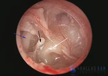
The posterior, or back part, of the eardrum is retracted into the middle ear cavity. The eardrum is sucked in considerably and is resting on the incus, the second hearing bone. Over time, this retraction onto the incus will cause erosion, or damage, to this hearing bone. The remainder of the eardrum appears to be in good health.
CO2 laser is utilized to tighten, or contract, the eardrum and to fix the area that is sucked in. The laser causes changes in the middle layer of the eardrum leading to fibrosis and thickening, and less chance of persistent retraction.
Severely Retracted Eardrum
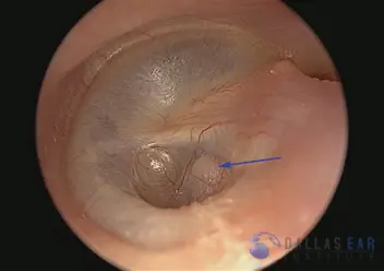
A severely retracted, or sucked in, eardrum is illustrated. This eardrum is extremely thin and lacks the structure of a normal eardrum. The facial nerve that travels through the middle ear is easily visualized because of the retraction of the eardrum (blue arrow). Over time, the pressure exerted by the eardrum on the bone that covers the facial nerve can cause resorption of this bone and exposure of the nerve.
The second hearing bone, the incus, has been eroded because of the retraction of the drum and is no longer present in the middle ear space. The third hearing bone, the stapes, is indicated by the black arrow. In a normal ear, this hearing bone is connected to the incus and this allows the transmission of sound. In this ear, the eardrum is completely sucked down onto the stapes and completely encases it and the bone that houses the inner ear (white arrow), like saran wrap. When the skin of the ear drum is draped on the hearing bones, significant erosion of the bones can occur and hearing loss and infection ensues.