Glomus Tympanicum Tumors
Cases Studies
Glomus Tumor Case Number 1
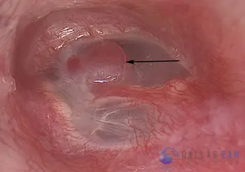
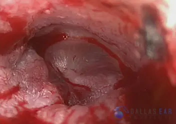
The eardrum is clearly visualized. There is a beefy-red mass underneath the eardrum that is actually touching the undersurface of the eardrum (black arrow). The red coloration of the mass is a function of the vascularity, or significant blood bloody supply, that this mass has. When viewed in real time, the mass actually pulsates and causes the eardrum to pulsate as well.
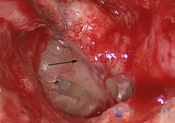
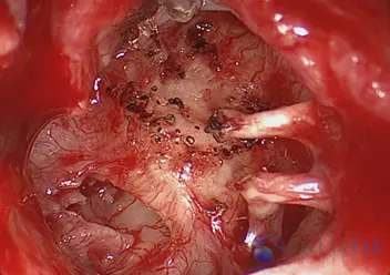
An incision has been made in the ear canal and the eardrum has been lifted up, exposing the middle ear cavity. The eardrum is lifted like the hood of your car, allowing full visualization of the inner ear. The dense bone of the inner ear is visualized. There are some scar bands from the inner ear bone to the glomus tumor, which originates from a blood vessel that lies on the solid bone of the inner ear (black arrow).
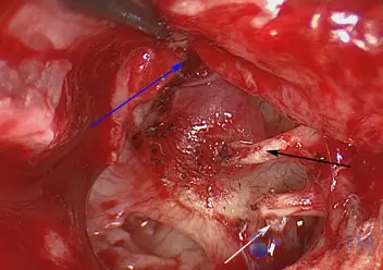
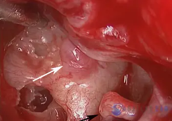
The eardrum has been lifted off of the malleus bone, which is the first hearing bone (black arrow). This allows complete visualization of the entire glomus tumor and will facilitate removal of the tumor. The blue arrow shows the undersurface of the eardrum that has been reflected forward. The white arrow shows the junction of the incus and stapes, the 2nd and 3rd hearing bones. These bones are not disturbed during this surgery.
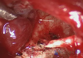
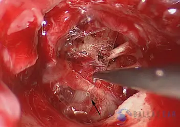
Using a CO2 laser, the very vascular tumor is peeled off of the bone of the inner ear. The laser facilitates removal because it allows the tumor to be removed without significant blood loss. The white arrow shows a feeding vessel to the tumor, which is controlled using the CO2 laser and, once again, limits the blood loss to miniscule amounts.
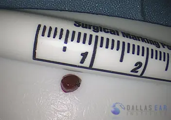
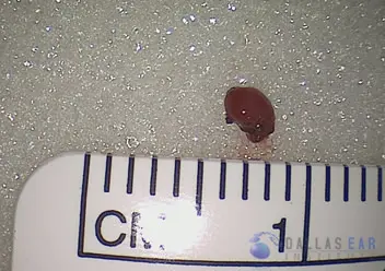
The middle ear tumor has been completely removed and is visualized on the back table. As you can see, its size is quite small (~4mm), though this takes up a significant amount of space within the middle ear cavity.
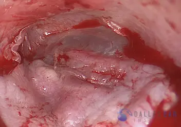
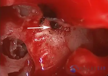
The tumor has been completed removed. The small black dots on the surface of the inner ear are areas where the tumor was removed using the assistance of the CO2 laser, ensuring no elements of the tumor remain behind.
A sheet of absorbable film is placed in the middle ear, between the eardrum and the inner ear bone, to prevent any scar tissue formation.
The eardrum is replaced and the ear canal is packed with a small amount of absorbable packing. The entire surgery is done through the ear canal, with no need for an incision behind the ear. The patient is sent home the same day, after recovering from the general anesthesia.
Glomus Tumor Case Number 2
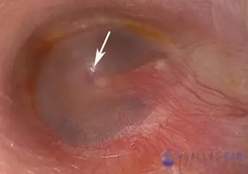
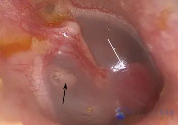
View of the eardrum through the ear canal. No incisions are made in the skin behind the ear—the approach is completely through the ear canal utilizing a high-magnification operating microscope. The eardrum is examined and there is a reddish mass seen in the middle ear cavity just inside of the eardrum. This is a glomus tympanicum tumor and it is just barely touching the eardrum.
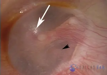
An additional view of the glomus tympanicum tumor within the middle ear, which is indicated by the white arrow. The black arrowhead is pointing towards one of the middle ear hearing bones that can be seen through the eardrum—this is the incus, the second hearing bone commonly called the “anvil” (hammer-anvil-stirrup).
The eardrum has been reflected up, much like the hood of a car is lifted. Inside of the eardrum, the contents of the middle ear are clearly visualized, including the incus and stapes hearing bones (black arrow). The white arrow indicates the glomus tympanicum tumor that originates from the promontory of the inner ear. This vascular tumor originates from cells surrounding blood vessels.
Using CO2 laser technology, the glomus tumor is removed. There is little bleeding and the excision is very precise because of the laser technology utilized. The origin of the tumor is also lased with the CO2 laser.
The eardrum is replaced. A slight amount of packing is used in the ear canal
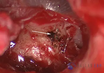
Glomus Tumor Case Number 3
The glomus tympanicum tumor is visalized in the middle ear behind the eardrum (white arrow). It is abutting the eardrum.
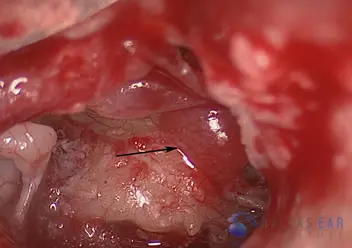
The eardrum has been elevated, exposing the middle ear space. The hearing bones are seen on the left of the image, and the glomus tumor is visualized (black arrow).
The tumor has been completely removed.
The tumor is removed with the assistance of CO2 laser, and the source of the tumor is treated with the laser to prevent the tumor from coming back in the future.