Stapedectomy Cases
Stapedectomy Case Number 1
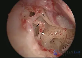
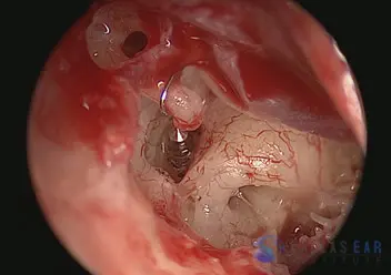
The eardrum has been lifted with an incision in the ear canal. The middle ear cavity is visualized. The malleus (black arrowhead), incus (black arrow), and stapes (white arrow) bone are clearly seen. These three bones make up the ossicular chain—the 3 middle ear bones that allow sound to be conducted into the inner ear. The chorda tympani nerve, one of the taste nerves, is seen traveling above the incus and just beneath the malleus bone.
The stapedius tendon, which connects to the stapes bone, is divided with the CO2 laser (black arrow).
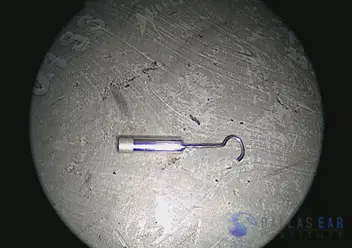
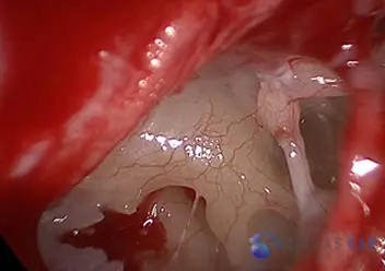
The stapes piston is prepared for insertion into the inner ear opening. This piston is 4.25 mm (less than half of a centimeter!) in length and 0.5 mm in width.
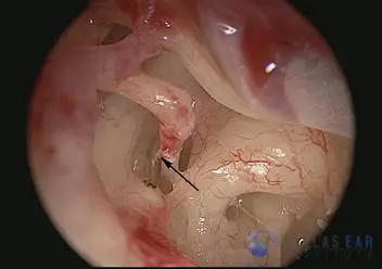
The stapes structure (stirrup) has been removed with the CO2 laser. The stapes footplate is easily seen now (black arrow). The footplate is the flat part of the stapes bone that is set into the oval window of the inner ear. This footplate separates the inner ear from the middle ear.
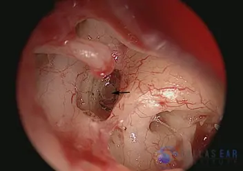
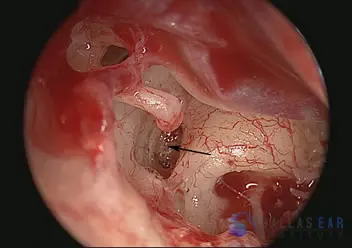
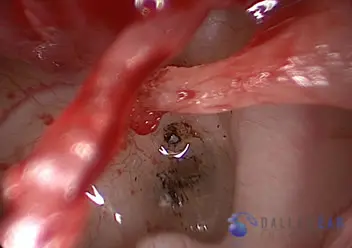
An opening has been created into the inner ear (black arrow) to allow placement of the prosthesis. The fluid of the inner ear is clearly visualized through the opening.
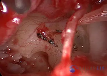
The piston hook is placed over the incus bone and the base inserted into the inner ear, recreating the hearing mechanism. The hook will be crimped over the incus bone. Now, as the eardrum moves with sound, the piston actually moves slightly in and out of the inner ear and allows the individual to hear.
Stapedectomy Case Number 1

The healthy ear drum is visualized through the ear canal.
Incision in the ear canal allows the ear drum to be elevated. The chorda tympani nerve is seen. The ossicular chain including the malleus, incus, and stapes is intact. However, the surgeon gently touches the stapes and confirms it is stuck.
The stapes is removed with a laser and the oval window is visualized.
The oval window is gently opened with a laser.
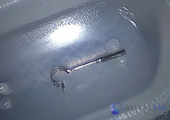
The stapes piston to be implanted is shown here.
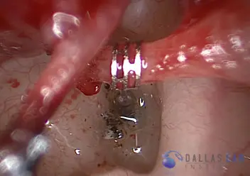
The piston is placed into the opening in the round window and hung on the incus.
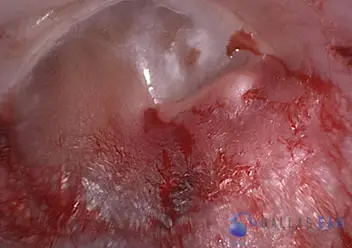
The ear drum is placed back into normal position and the procedure is complete.